Next Step: Make Cards on the Automatic Key Concepts, and Vignettes
Remember, the more you automatically know what each sentence means on your test, the better you will do. There are 4 stages in making interpretation more automatic:
- Stage 1: Unable to Make Pathophysiologic Chronologies in Either Timed or Untimed setting
- Stage 2: Basic Pathophysiologic Chronologies, but with Significant Gaps
- Stage 3: Detailed Pathophysiologic Chronology Without Time, but Unable to Consistently Generate PC During Timed Setting
- Stage 4: Consistent Pathophysiologic Chronologies in Timed Setting
My goal with these vignettes is to help you reach Stage 4. How do you do so?
- With the Automatic Key Concept cards, you can master the underlying information to move past Stages 1 + 2.
- Then, with the Vignette/Pathophysiologic Chronology cards, you can teach yourself to make these connections on your exam.
Automatic Key Concepts:
Copy + paste these into your cards, to make these key concepts more automatic.
How is nuclear medicine different from X-rays/ CT scans?
Nuclear medicine: radioactive material enters the body, then decays, and emits electromagnetic radiation from within the tissues of interest.
X-rays / CTs: x-ray radiation is shot at the body, and is detected on the other side by a detector.
What does tomography mean?
Imaging by sections or sectioning. through the use of any kind of penetrating wave.
What are the 3 major kinds of nuclear medicine scans?
PET(positron emission tomography), scintigraphy, and SPECT (single-photon emission computed tomography)
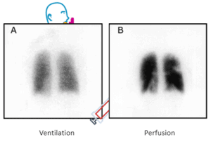
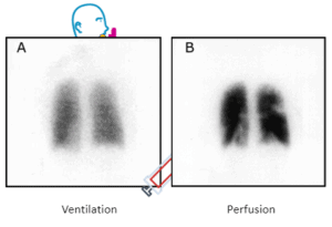
A V/Q scan is an example of nuclear medicine. How are the different ventilation and perfusion images obtained?
Patient given an inhaler, with radioactive material. Inhaled material distributes to the lungs, and as it decays, the radiation is picked up by a detector. Then, the patient is given an IV injection of (different) radioactive material, which distributes to the lungs, and as it decays, will emit electromagnetic radiation that is detected.
What kind of nuclear medicine scan is V/Q? (Scintigraphy vs. SPECT vs. PET)
Scintigraphy (2D, using gamma radiation)
In PET scans, the radioactive material is bound to what substance? Why does this make sense?
Bound to glucose-cancer often consumes a lot of glucose, and so the cancer will take up extra glucose (and thus emit extra electromagnetic radiation) relative to surrounding tissues, Cancer consumes extra glucose because it often has decreased mitochondrial activity, and undergoes less aerobic metabolism. As such, cancer cells often rely on anaerobic glycolysis, which requires much more glucose than aerobic metabolism. (For more, see: “Biochemistry 1: TCA Cycle, ETC”)
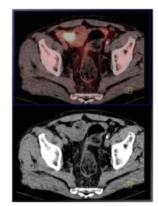
For the included images, what are the images above and below? How were they obtained?
Above: Pet scan, overlaying a CT scan
Below: CT scan
The PET and CT scans are obtained separately. (PET scans work by injecting glucose that is bound to radioactive material-because cancer uses glucose more than surrounding tissues, it is oftern “brighter” on a PET scan)
Using the pathophysiology, explain the presentation of stable angina
Fixed coronary artery stenosis → under physiologic stress (exercise, emotional upset, etc.), cardiac demand > coronary artery supply → ischemia → pain during exertion, but no pain at rest
What is the basis for a stress test? What are you looking for?
Because for a fixed coronary stenosis, blood supply at rest may be sufficient to meet myocardial demand. However, only during physiologic stress (e.g., exercise), when myocardial demand ↑, you will see ischemia. Thus, stress tests are designed to increase myocardial demand, to assess for coronary stenosis that would lead to ischemia only during rest.
Exercise vs. chemical stress test: why would you do one vs. the other?
Exercise stress tests are usually the default. However, in the case someone can’t exercise (e.g., movement limited by pain, bad hips, etc.) → chemical stress test.
Why do people typically get stress tests? Why did our patient in the vignette get one? (55-year old man with type 2 diabetes mellitus, normocytic anemia, and chronic kidney disease with exertional dyspnea)
Often to assess for coronary artery disease as the cause of a nonspecific symptom/sign. For example, chest pain/dyspnea can occur for a variety of reasons, one of which is coronary artery disease. In this case, this patient had risk factors for CAD, and so to assess their exertional dyspnea, a stress test was ordered.
Vignette/Pathophysiologic Chronologies:
Copy + paste these into your cards, to make the connections behind these vignettes more automatic.
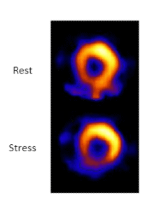
A 55-year-old man with type 2 diabetes mellitus, normocytic anemia, and chronic kidney disease comes to see his primary care physician for evaluation of exertional dyspnea. Blood pressure is 141/93 mmHg, and pulse is 82/min. BMI is 35 kg/m2. Single photon emission CT images at rest and after 5 minutes of exercise on a treadmill, using separate intravenous injections of technetium-99-labeled perfusion agent are shown to the side.
What is the pathophysiologic chronology?
What is the significance of the images of “stress” and “rest” images?
Summary:
Coronary artery disease → stable angina → to pain/ischemia under physiologic stress → reversible ischemia on nuclear medicine scan.
Detailed:
Born healthy → cardiovascular risks factors → develop stable plaque in coronary artery → occlusion > 70% of lumen → stable angina/ → ischemia during physiologic exertion → during stress portion of nuclear medicine scan develops ischemia as demand > supply


